- Feel Free to contact us
- +886 4 2276 6193
- info@duogenicsc.com

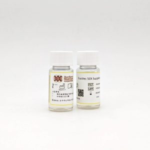
DuoVEC is a xeno-free supplement optimized for human endothelial cells (hEC) culture.
Human umbilical vein endothelial cells (HUVEC) culturing in DuoVEC express typical EC protein markers throughout P1 to P7 and the cell doubling time < 40 hours.
Storage : -15°C or below, protect from light.
Shelf Life : 12 months
Avoid freeze / thaw cycles
| Product Name | Catalog Number | Size | Grade |
| DuoVEC – Supplement 50X | DVM500-S | 10mL | Research Use Only |
Research in endothelial cells have always been a hotspot in biomedical area for the following reasons: cardiovascular disease is the leading cause of death worldwide; angiogenesis plays a curtail part in tumor formation; and due to the shortage of corneal donation. It’s known that ECs take part in blood pressure regulation, arteriosclerosis, angiogenesis, coagulation and so on. HUVEC provide a classic model system to study many aspects of endothelial function and disease, including cardiovascular and metabolic diseases. This model is still widely used today mainly because of the high HUVEC isolation success rate.

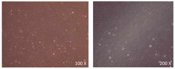
The umbilical cord surface was washed with IMDM (or PBS), and the same medium was injected inside the umbilical vein by syringe to remove the clots. IMDM with 2 mg/ml collagenase NB 4(SERVA) was then injected into the umbilical vein and left for 30 minutes at room temperature. The medium was then squeezed out from the umbilical cord and centrifugate at 15,000 rpm for 5 minutes. The cells were seeded on a 100 mm TC treated culture plates pre-coated with 10 ug/ml fibronectin. The pictures were taken 5 days after isolation.

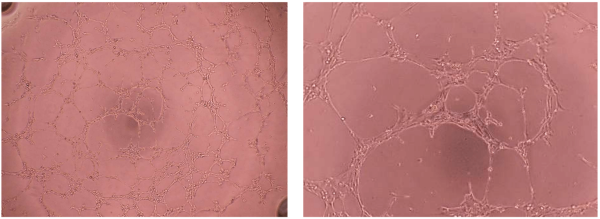
HUVEC were isolated and cultured for 1 passage prior to this experiment. Cells were then seeded on 96 well culture plate pre-coated with 100 ul Matrigel. And the cells were left in a 37°C, 5% CO₂ incubator for at least 4 hours before the pictures were taken.

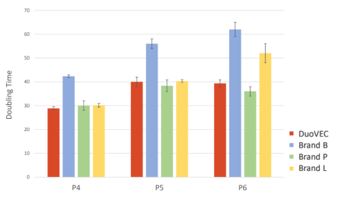
Doubling time of HUVEC (from P4 to P6) culturing in :
1. DuoVEC
2. Brand B XF medium
3. Brand P FBS medium
4. Brand L FBS medium
HUVEC grow better in DuoVEC than in Brand B Xeno-Free medium.
DuoVEC provided competitive growth rate when culturing HUVEC compare to FBS medium.

Flow cytometry analysis of HUVEC cultured in DuoVEC at P1 and P7. CD31 and CD144 are both highly expressed.
User manual and more information are available upon request.
info@duogenicsc.com